A focused ultrasound that measures the cervix to identify pregnancies at risk of preterm birth, while also evaluating placental position and screening for vasa previa.
Timing
16–24 weeks
Length
< 2 minutes
Threshold
25 mm
01Why transvaginal
A clearer view, where it matters.
01 · The threshold
The millimeter that changes the plan.
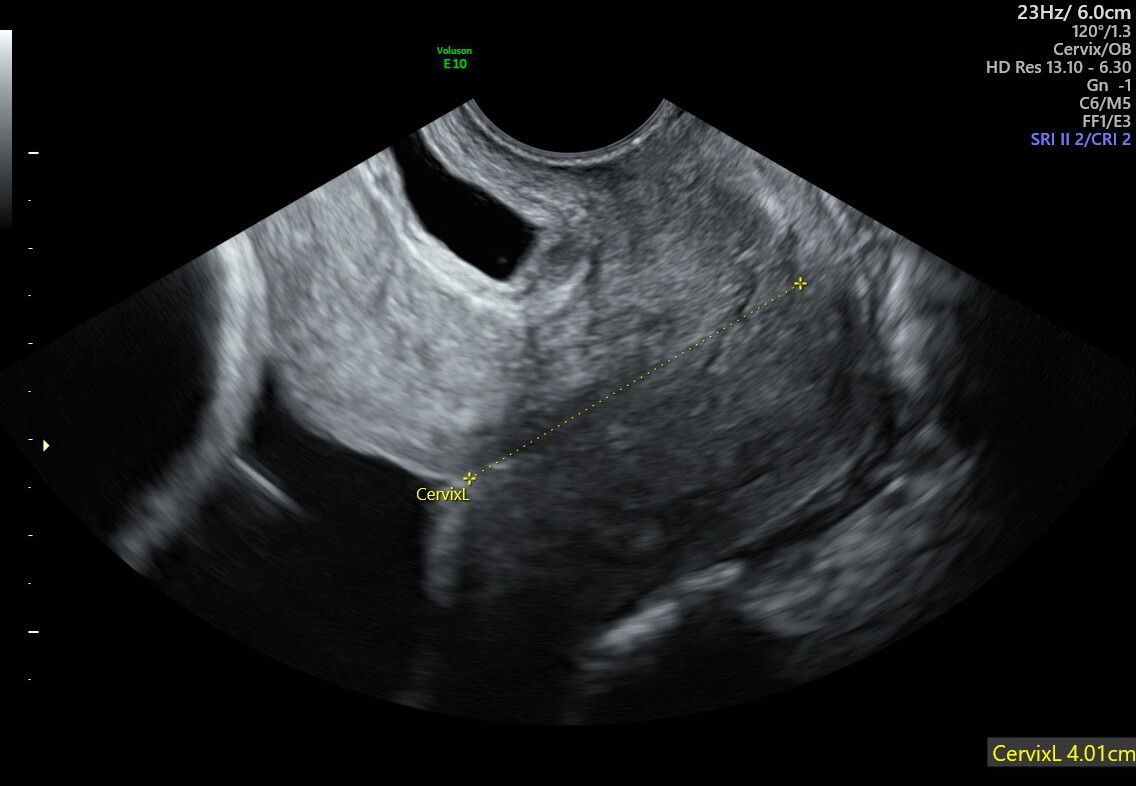
A cervical length above 25 mm is reassuring. Below it, we partner with your OB on a focused follow-up plan to help keep your pregnancy on track.
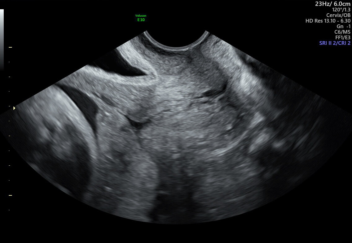
A transvaginal ultrasound uses a slender probe placed in the vaginal canal, so the cervix can be measured directly without the abdominal wall, bladder, or surrounding tissue blurring the image.
Resolution
Closer proximity allows higher-frequency sound waves and a much sharper image of the cervix.
Precision
For cervical length, even a few millimeters can change a clinical decision. This is the gold standard.
Earlier signal
Subtle structural changes in the second trimester are simply not visible from the outside.
Safe in pregnancy
Transvaginal ultrasound uses the same harmless sound waves as an abdominal scan. There is no radiation, and the exam is considered safe at every stage of pregnancy. It will not cause bleeding, infection, or harm to your baby.
This technique has been used safely for decades and is recommended by both the American College of Obstetricians and Gynecologists and the Society for Maternal-Fetal Medicine as the gold standard for evaluating cervical length and placental position.
02Cervical length screening
The gatekeeper of your pregnancy.
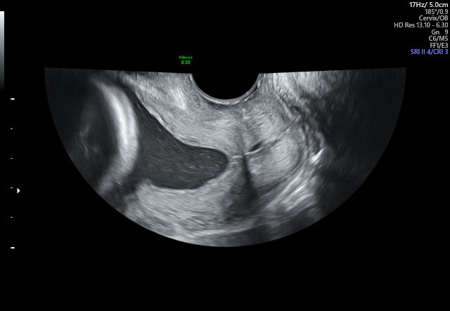
The cervix is the lower part of the uterus. In a healthy pregnancy it stays long, thick, and closed until your baby is ready to be born. If it begins to shorten, thin, or open too early, the risk of spontaneous preterm birth goes up.
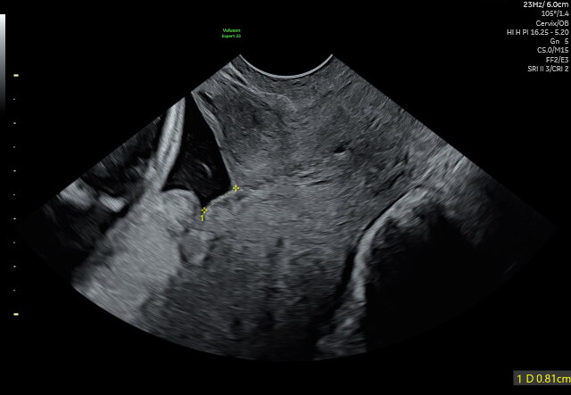
We typically look for a cervical length greater than 25 millimeters between 18 and 24 weeks. We also watch for funneling, when the inner part of the cervix begins to open into a V or U shape even while the outer part stays closed.
Reassuring
Normal cervix
Long, thick, and closed. The internal os stays sealed throughout pregnancy.
Follow-up needed
Funneling cervix
The internal os opens into a V or U shape, while the external os may still appear closed.
Who benefits from screening
Everyone — including patients with no risk factors at all.
Cervical shortening can occur even in patients with no prior risk factors or warning symptoms. Because of this, many experts support evaluating cervical length during the mid-trimester anatomy ultrasound (18–24 weeks) as part of routine pregnancy care.
Especially important for
A prior spontaneous preterm birth
Twins or higher-order multiples
Pelvic pressure, cramping, or other symptoms
A history of cervical surgery
A second-trimester pregnancy loss
No known risk factors at all
Measuring cervical length is simple, safe, and helps identify pregnancies that may benefit from early treatment to reduce the risk of preterm birth. Early detection allows us to act early — often before symptoms appear.
When we find a short cervix
03Placenta previa
How close is the placenta to the exit?
The placenta delivers oxygen and nutrients to your baby. Where it sits inside the uterus matters for how you will deliver. Placenta previa is when the placenta covers all or part of the cervical opening, which can cause heavy bleeding during a vaginal delivery.
Transvaginal imaging gives a clear, unobstructed view of the relationship between the placental edge and the cervix, so the diagnosis is accurate. An abdominal scan can sometimes give a false positive when the bladder is full or the lower uterus is contracting.
Low-lying
Within 2 cm
The edge of the placenta sits close to the cervix but does not cover it.
Complete previa
Covering the cervix
The placenta entirely covers the internal opening, requiring a planned cesarean.
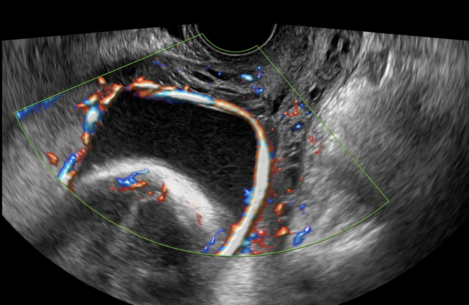
04Vasa previa
A rare but serious condition we look for every time.
Vasa previa is a rare but very serious condition where unprotected fetal blood vessels run across the cervical opening. If those vessels rupture during labor or when the water breaks, it can cause rapid, life-threatening blood loss for the baby.
During your exam we use Color Doppler imaging, which shows blood flow in real time, to sweep the area over the cervix and confirm that no fetal vessels are crossing it. When we do find vasa previa, we build a specialized care plan with your OB, usually a scheduled cesarean before labor begins.
Color Doppler
Lets us see blood flow in real time, appearing as red and blue movement on the screen, so any vessel crossing the cervix is impossible to miss.
05Your comfort
What to expect during the exam.
We know a transvaginal ultrasound can feel more personal than an abdominal scan. Here is exactly what happens, step by step, so you arrive knowing what to expect.
01
Privacy
A private room to change, with a sheet for coverage.
02
The probe
Slender, sterile sheath, warm gel, much thinner than a speculum.
03
Sensation
Mild pressure, similar to a pelvic exam. Pause anytime.
04
Under 2 minutes
Brief and focused. Most patients find it goes by quickly.
Before you arrive
A transvaginal scan is best performed with an empty bladder. Please use the restroom before your exam begins.
Common questions
Frequently asked.
Don't see your question? Call our office and we will be happy to walk you through anything before your appointment.
It should not be painful. Most patients describe a mild pressure, similar to a pelvic exam. The probe is slender, well lubricated, and you can ask us to pause or adjust at any time.
A full bladder lifts the uterus away from the probe and obscures the view. For transvaginal imaging, an empty bladder lets the probe sit close to the cervix and produce the sharpest image.
Yes. Like all ultrasound, it uses sound waves rather than radiation. It is the standard of care for evaluating the cervix and lower uterus during pregnancy.
It depends on your history and what we find. Some patients need a single screening. Others, particularly with a prior preterm birth or a short cervix, may need follow-up scans every one to two weeks during the second trimester.
Ready to schedule your screening?
Most patients arrive with a referral from their OB. If you don't have one yet, we can help coordinate. Same-week appointments are usually available.